TS-PScan
TS-PScan
Research Use Only
Live
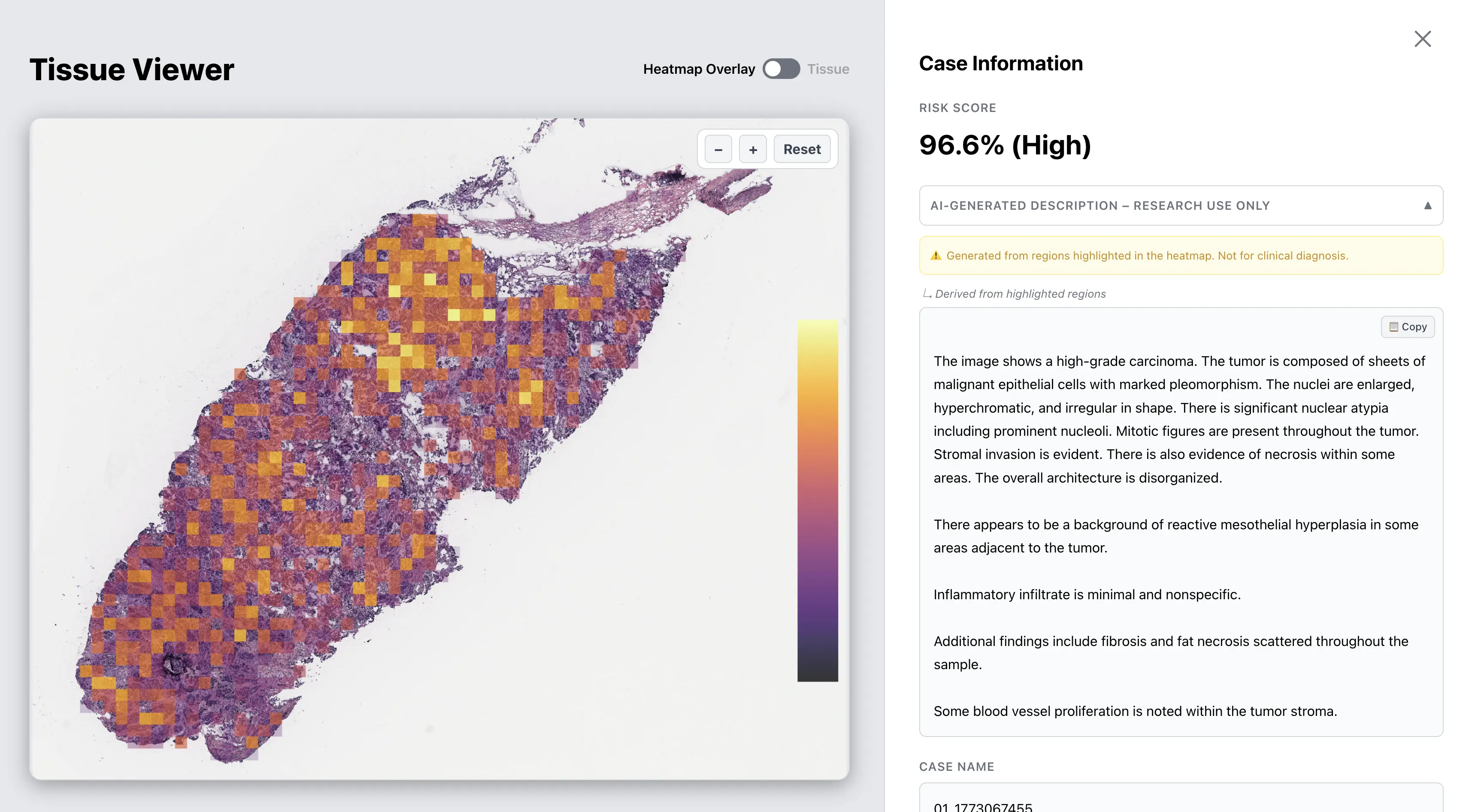
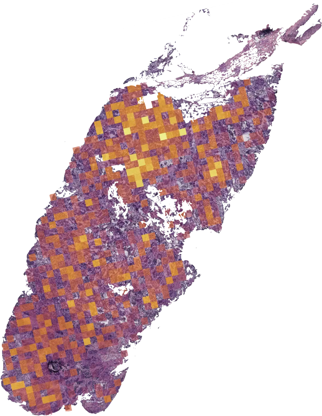
Pan-cancer screening tool. Classifies H&E whole-slide images as cancer or normal across 11 tissue types. Outputs a calibrated risk score and attention heatmap. Available for research access upon request.